2025: Volume 5, Issue 6
Past Issues
 Abstract
  PDF
PDFLong-Term Experience with 1064-nm Nd: YAG Q-Switched Laser Treatment for Onychomycosis
Hector Ricardo Galvan-Garcia1,*, Julio Cesar Aguilar-Perez2, Andrea Isabel Mendez-Juarez2, Maria del Pilar Alvarez-Alatriste3, Lopez Garcia Citlally Nayelli4
1Dermatologist at Dermoquirúrgica gg clinic. Guadalajara, Jalisco, Mexico
2Dermatology Resident, Dr. José Barba Rubio Dermatological Institute of Jalisco, Zapopan, Jalisco, Mexico
3Internal Medicine Resident, IMSS BIENESTAR Tampico Specialty Hospital, “Dr. Carlos Canseco,” Tampico, Tamaulipas, Mexico
4Internal Medicine Resident, National Medical Center of Bajío UMAE T1 IMSS. León, Guanajuato, Mexico
*Corresponding author: Hector Ricardo Galvan-Garcia, Dermatologist at Dermoquirúrgica gg clinic. 2261 López Cotilla. 44130, Guadalajara. Jalisco, Mexico, Email: [email protected]
Received Date: July 17, 2025
Publication Date: October 07, 2025
Citation: Galvan-Garcia HR, et al. (2025). Long-Term Experience with 1064-nm Nd: YAG Q-Switched Laser Treatment for Onychomycosis. Dermis. 5(6):52.
Copyright: Galvan-Garcia HR, et al. © (2025).
ABSTRACT
Introduction: laser treatment for onychomycosis has been described as an easy to perform and safe method. Objective: to evaluate the efficacy and safety of 1064-nm neodymium-doped yttrium aluminum garnet (Nd:YAG) Q-switched laser for the treatment of onychomycosis. Method: This retrospective study included 700 patients with suggestive onychoscopy. KOH direct examination confirmed diagnosis of onychomycosis of patients who were treated with 1064-nm Nd:YAG Q-switched laser from April 2012 to December 2023. Results: Most of the patients required only 1 session (86%) and showed clinical response at 6 months while 14% required a second laser session. Clinical and mycological response was seen in 100% at 12 months. There were no documented side effects. Conclusions: Our long-term experience confirmed the efficacy and safety of Nd:YAG Q-switched laser treatment of onychomycosis.
Keywords: Onychomycosis, Nd:YAG laser.
INTRODUCTION
Onychomycosis is a common disease with a prevalence between 2% to 28% with high recurrence rates [1,2]. The most common pathogens are the dermatophytes Trichophyton rubrum (T. rubrum) and T. mentagrophytes, in 60%-90% of the cases, less common pathogens are other dermatophytes, molds and yeasts [1,2]. Onychomycosis affects fingernails and toenails; however, toenail involvement is more common. The clinical appearance is suggestive and can be supported by dermoscopy, an inexpensive and quick tool. The dermoscopic features of onychomycosis are ruin appearance (ventral indentations of the nail plate caused by dermal debris), subungual hyperkeratosis, longitudinal striae, onycholysis, distal irregular termination (distal pulverization of the nail plate), chromonychia (multicolored pigmentation), leukonychia and melanonychia [3].
The diagnosis is not only clinical since mycologic laboratory testing is necessary. Direct examination with 5%-40% KOH is the preferred diagnostic tool, it has a sensitivity of 61% (44%-100%) and specificity of 95% (75%-100%) and the report takes only minutes to hours, with the disadvantages that it does not identify the pathogen and is operator dependent. Fungal culture has a sensitivity of 56% (29%-82%) and specificity of 99% (83%-100%) but takes two to four weeks to grow. Lastly, PCR has a sensitivity of 85-100% and specificity of 94%-100% with the disadvantage of higher cost [2].
Current treatments for onychomycosis comprehend topical or oral antifungal medication. Topical treatments have limited efficacy, and oral antifungals are highly effective due to their capability of penetration in nail plates, however they might cause adverse events such as hepatic and renal damage. Considering all this, other therapeutic options have been sought for [1,4].
Laser therapy was approved for onychomycosis in 2010 [1], its mechanism of action is thought to be by its activity on the pigment particles or chromophore groups of the fungal cells by increase of the temperature, thereby inhibiting their growth [5]. Furthermore, it has the benefit of causing few side effects, like mild and limited erythema and swelling after the procedure [6].
Objective
The objective of our study was to assess the effectiveness, time for resolution, minimum of sessions needed to achieve clinical and mycological clearance and safety of the 1064-nm Nd:YAG Q-switched laser treatment of onychomycosis.
METHODS
After searching through the database of the clinic, 700 patients were found and included in the study from April 2012 to December 2023. All patients had a KOH-confirmed diagnosis of onychomycosis. Patients of all ages who had not previously received any type of treatment were included, each patient signed a consent form allowing for clinical photographs of their nails to be taken before and after the laser therapy. Pregnant women, patients with subungual hematoma, taking photosensitizing drugs and those affected by psoriasis, lichen planus or atopic dermatitis with nail involvement were excluded.
Dermoscopy was performed in each patient. The most frequent signs were ruin appearance, longitudinal whitish striae, subungual hyperkeratosis, chromonychia and melanonychia (Figure 1). A direct microscopic test was performed with a mix of 25% KOH and 5% glycerol (1 hour at 51-54ºC) for lipid emulsification, and mycological structures were identified under ´3400 magnification. This test was performed at the beginning and at the end of the evaluation for each patient.
Laser therapy was performed with 1 of 3 1064-nm Nd:YAG Q-switched laser devices: 1) Monalisa Laser, Sincoheren Co., Xizhimen Beidajie, Beijing, China, 2) Q-Clear TM, Light Age, Inc., Somerset, NJ, USA, and 3) Discovery Pico Quanta System, Milano Italy. Laser settings were adjusted to deliver a fluency of 500-800 mJ/cm2 (4-6 J/cm2 equivalent) on a 3 mm spot at 5-10 Hz nanoseconds, in a single session. Three applications were performed on a square configuration across the entire ungual plate for each case. Immediate changes after laser light application were observed and determined as nail clearance. Treatment questionnaires and clinical revisions were scheduled at 3, 6, 9, 12 and 18 months after treatment.
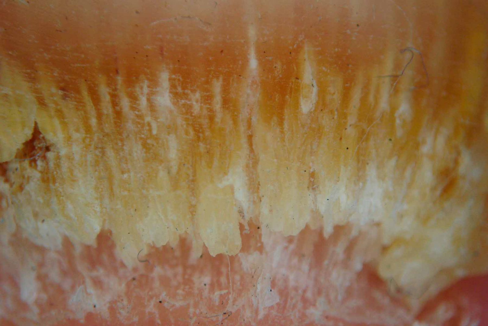
Figure 1. Dermoscopic findings: ruin appearance and longitudinal striae.
RESULTS
Seven hundred patients were included in the clinical study, 670 adults and 30 children. Ages ranged between 6 and 79 years. A total of 2800 nails were treated: 2600 toenails and 200 fingernails; twenty patients had both toenails and fingernail involvement. Clinical response was seen in 84% of the patients within 3 months of the laser treatment with proximal healthy grow of the nail (Figure 2). At 6 months 86% showed clinical response and 14% required a second laser session considering the lack of visible proximal healthy grow. At 12 months, all the patients (100%) showed clinical and mycological response with negative KOH test (Figure 3). There were no side effects at all.
During laser therapy, we observed near instantaneous “clearance” (change in the appearance from dark to clear) of the affected nails, particularly nails that showed melanonychia or dark green pigmentation. Additionally, as the clearance was evident the characteristic cracking sound of the fungus-affected areas of the ungual plate diminished until cessation, similar to what happened when laser treatment was administered to the nearby clinically healthy zones.

Figure 2. Clearance line response at 3 months after treatment.
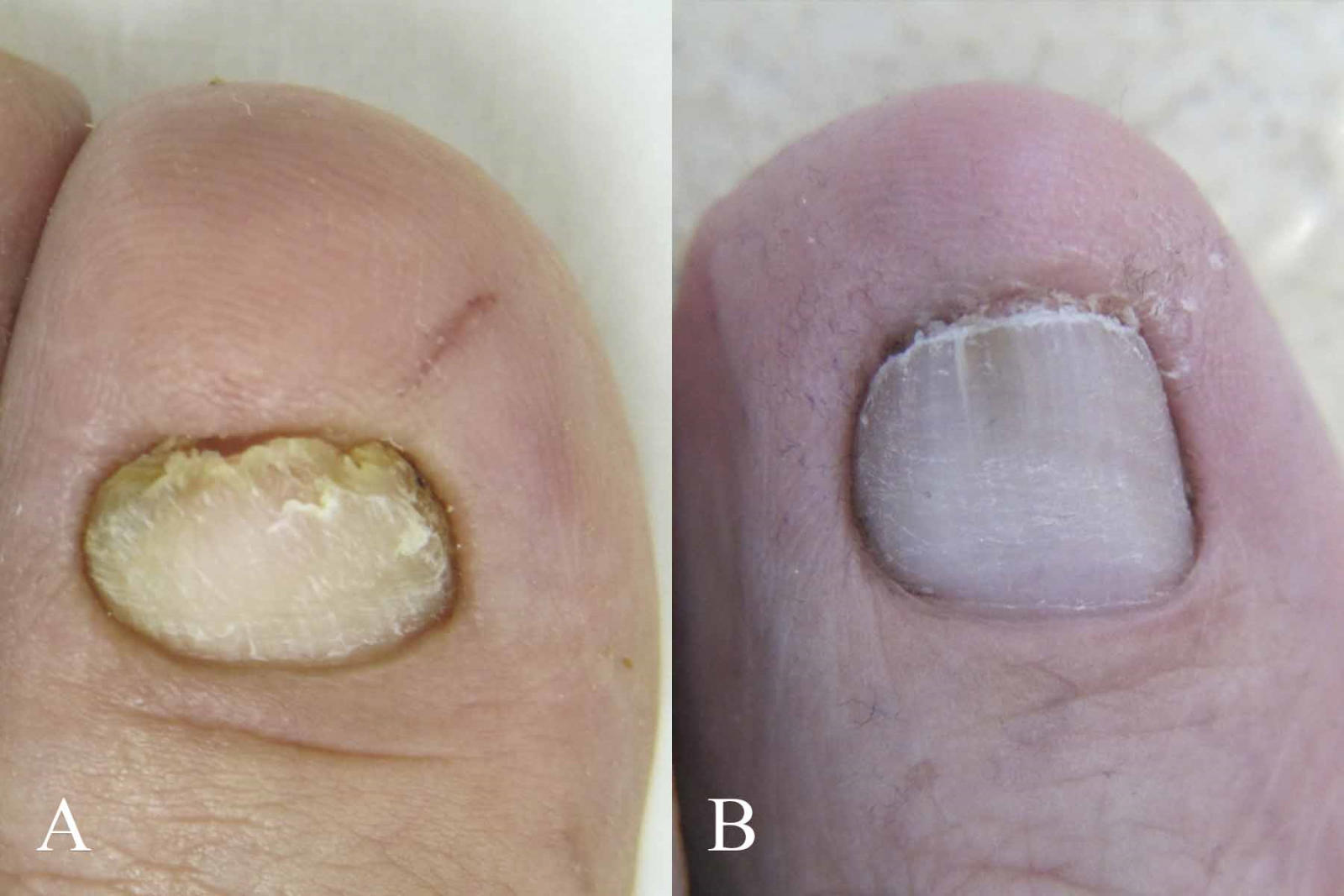
Figure 3. (A, B) Comparison of toenails showing complete response after 12 months of treatment.
DISCUSSION
Cao Y, et al. [1], confirmed that long-pulsed 1064-nm Nd:YAG laser is effective for treating onychomycosis, and the energy irradiation can change cellular ultrastructure and inhibit colony growth of T. rubrum and T. mentagrophytes. The Nd:YAG Q-switched laser has been described under this name because it generates high-energy peaks at many repetitions. It produces energy that mechanically damages only the target of interest through selective photothermolysis on the pigment of fungi structures and it does not warm the tissue, so it does not elicit pain.
In a study of Meretsky CR, et al. [4], laser treatment was effective for onychomycosis compared to terbinafine and other antifungals, additionally, mycological cure rates were higher among participants receiving laser therapy as compared to terbinafine (OR = 3.19, 95% CI: 1.39-7.29; p = 0.05) with fewer adverse effects. However, the evidence was based on small comparative studies. In the present study a significant number of patients were treated with Nd:YAG Q-switched laser therapy alone showing efficacy at 12 months with both clinical and mycological cures.
Laser therapy has been compared with itraconazole pulses, with similar mycological and dermoscopic cure rate with no statistical difference [7]. A combination of long pulsed Nd:YAG laser and itraconazole pulse therapy gives best clinical results when compared to itraconazole pulse therapy alone [8]. On the other hand, in the study conducted by Rovers JFJ, et al. [9], after 3 sessions with Nd:YAG laser 1064 nm, only 12.4% of patients had total clinical clearance, and the study conducted by Wanitphakdeedecha R, et al. [6], demonstrated mycological clearance in 40.7% after 1 month (4 sessions) of Nd:YAG. Nevertheless, and as stated in our results, longer follow up time is needed to assess both clinical and mycological cures and to determine if multiple sessions are required, since the nail plate of the toenails can take from months to over a year to fully grow new.
The greatest advantages of laser treatment for onychomycosis compared with traditional treatments such as oral or topical medications are the easy application, scarce contraindications and overall absence of side effects.
CONCLUSIONS
The present study confirmed the efficacy and safety of 1064-nm Nd:YAG Q-switched laser therapy as a sole and an uncomplicated treatment for onychomycosis.
ACKNOWLEDGEMENTS
None.
CONFLICTS OF INTEREST
The authors declare no conflicts of interest.
REFERENCES
- Cao Y, Xu S, Kong W, Xu Y, Fang H. (2020). Clinical retrospective analysis of long-pulsed 1064-nm Nd:YAG laser in the treatment of onychomycosis and its effect on the ultrastructure of fungus pathogen. Lasers Med Sci. 35(2):429-437.
- Falotico JM, Lipner SR. (2022). Updated Perspectives on the Diagnosis and Management of Onychomycosis. Clin Cosmet Investig Dermatol. 15:1933-1957.
- Litaiem N, Mnif E, Zeglaoui F. (2023). Dermoscopy of Onychomycosis: A Systematic Review. Dermatol Pract Concept. 13(1):e2023072.
- Meretsky CR, Friday BL, Schiuma AT. (2024). Efficacy of Laser Therapy in Comparison With Other Methods for the Treatment of Onychomycosis: A Systematic Review and Meta-Analysis. Cureus. 16(5):e59720.
- Vural E, Winfield HL, Shingleton AW, Horn TD, Shafirstein G. (2008). The effects of laser irradiation on Trichophyton rubrum growth. Lasers Med Sci. 23(4):349-353.
- Wanitphakdeedecha R, Thanomkitti K, Bunyaratavej S, Manuskiatti W. (2016). Efficacy and safety of 1064-nm Nd:YAG laser in treatment of onychomycosis. J Dermatolog Treat. 27(1):75-79.
- Nasif GA, Amin AA, Ragaie MH. (2023). Q-switcheded Nd:YAG laser versus itraconazole pulse therapy in treatment of onychomycosis: A clinical dermoscopic and mycologic study. J Cosmet Dermatol. 22(6):1757-1763.
- Hamed Khater M, Khattab FM. (2020). Combined long-pulsed Nd-Yag laser and itraconazole versus itraconazole alone in the treatment of onychomycosis nails. J Dermatolog Treat. 31(4):406-409.
- Rovers JFJ, Wagter LV, Greijmans EGE, Bovenschen HJ. (2021). 1064-nm Nd:YAG laser treatment for onychomycosis: is it worthwhile? Lasers Med Sci. 36(2):463-467.